

| Typical study duration | 6–8 hours of recorded sleep (American Academy of Sleep Medicine guidelines) |
| Number of channels recorded | Up to 20+ simultaneous physiological signals (Standard PSG protocol) |
| AHI threshold for OSA diagnosis | ≥5 events per hour (with symptoms) or ≥15 events per hour (AASM diagnostic criteria) |
| Normal REM proportion | Approximately 20–25% of total sleep time (Established sleep architecture norms in healthy adults) |
| Normal slow-wave sleep (N3) proportion | Approximately 13–23% of total sleep time in adults (Established sleep architecture norms) |
| Home sleep apnea test vs. PSG | Home tests measure fewer channels; PSG required for complex cases (AASM clinical practice guidelines) |
What Polysomnography Is — and Why Clinicians Order It
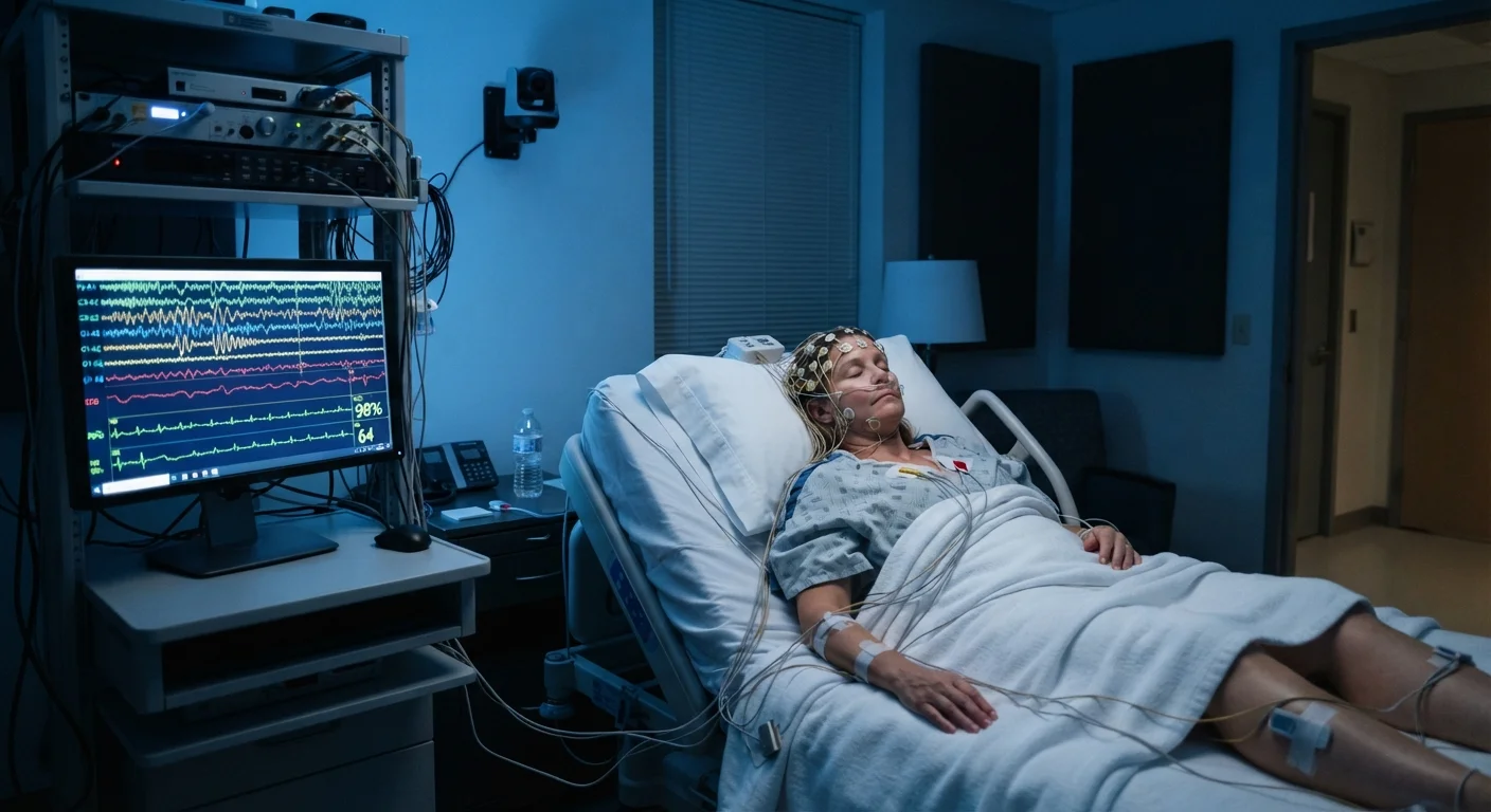
Polysomnography (PSG) is the clinical gold standard for diagnosing sleep disorders. Conducted overnight in an accredited sleep laboratory, it simultaneously records multiple physiological signals while you sleep — giving clinicians a layered, objective picture of your sleep physiology that no wearable or questionnaire can replicate.
A physician may order a PSG when evaluating suspected obstructive sleep apnea, narcolepsy, REM sleep behavior disorder, periodic limb movement disorder, or unexplained excessive daytime sleepiness. Understanding what each sensor actually measures helps you interpret your own results with greater confidence.
For a broader look at how sleep stages unfold throughout the night, see the architecture of a night's sleep.
Polysomnography (PSG)
A comprehensive overnight sleep study that simultaneously records brain activity, eye movements, muscle tone, heart rhythm, respiration, and blood oxygen levels. It is considered the clinical gold standard for diagnosing sleep disorders.
Apnea-Hypopnea Index (AHI)
A composite metric expressing the average number of complete breathing pauses (apneas) and partial reductions in airflow (hypopneas) per hour of sleep. Clinicians use AHI to grade the severity of sleep-disordered breathing.
Sleep Efficiency
The ratio of total sleep time to total time spent in bed, expressed as a percentage. It indicates how consolidating a person's sleep is relative to the opportunity available.
Muscle Atonia
The temporary paralysis of voluntary skeletal muscles that normally occurs during REM sleep. It prevents people from physically acting out dream content.
Oxygen Desaturation Index (ODI)
The number of times per hour that blood oxygen saturation drops by a specified threshold (commonly 3% or 4%) below baseline. ODI complements AHI in characterizing sleep-disordered breathing severity.
Arousal Index
The average number of brief EEG arousals — momentary shifts toward lighter sleep or wakefulness — per hour of sleep. A high arousal index reflects fragmented, non-restorative sleep.
The Core Measurements Explained
Electroencephalography (EEG) — Electrodes placed on the scalp record the electrical activity of the brain's cortex in real time. Distinct wave patterns mark each sleep stage: high-frequency, low-amplitude beta waves during wakefulness; slower alpha waves during relaxed wakefulness; theta waves in light sleep (N1); sleep spindles and K-complexes in N2; and the large, slow delta waves that define deep slow-wave sleep (N3). REM sleep produces a low-amplitude, mixed-frequency pattern resembling wakefulness. EEG is the primary signal used to score sleep stages.
Electrooculography (EOG) — Electrodes near the outer corners of the eyes capture eye movement. The hallmark slow rolling eye movements of N1 and the rapid eye movements of REM sleep are both identified here. EOG data is essential for confirming REM onset and ruling out sleep-stage misclassification.
Electromyography (EMG) — Surface electrodes on the chin and legs measure muscle tone. Muscle atonia — the suppression of voluntary muscle activity — is a defining feature of healthy REM sleep and its absence can signal REM sleep behavior disorder. Leg EMG channels detect periodic limb movements, which can fragment sleep without the sleeper's awareness.
Electrocardiography (ECG/EKG) — A single-lead ECG monitors heart rhythm throughout the study. This allows technicians to identify arrhythmias that may coincide with apneic events or occur independently during sleep.
Pulse Oximetry — A finger probe measures blood oxygen saturation (SpO₂) continuously. In obstructive sleep apnea, repeated airway collapses cause oxygen desaturations — dips below baseline that the scoring physician uses to calculate the oxygen desaturation index (ODI).
Respiratory Airflow and Effort — A nasal-oral thermistor or pressure transducer detects airflow at the nose and mouth. Elastic bands around the chest and abdomen record breathing effort. Together, these signals distinguish obstructive apneas (effort present, airflow absent) from central apneas (both effort and airflow absent) and from hypopneas (partial reductions in airflow).
Body Position Sensor — A positional sensor records whether you are supine, prone, or lateral. Many people experience positionally dependent sleep apnea, with apnea severity worsening significantly when lying on the back.
Video and Audio Recording — Infrared video and sound recording allow technicians to observe behaviors — sleepwalking, sleep talking, snoring, or unusual movements — that correlate with the physiological data.
Key Metrics Your Report Will Reference
| Typical study duration | 6–8 hours of recorded sleep (American Academy of Sleep Medicine guidelines) |
| Number of channels recorded | Up to 20+ simultaneous physiological signals (Standard PSG protocol) |
| AHI threshold for OSA diagnosis | ≥5 events per hour (with symptoms) or ≥15 events per hour (AASM diagnostic criteria) |
| Normal REM proportion | Approximately 20–25% of total sleep time (Established sleep architecture norms in healthy adults) |
| Normal slow-wave sleep (N3) proportion | Approximately 13–23% of total sleep time in adults (Established sleep architecture norms) |
| Home sleep apnea test vs. PSG | Home tests measure fewer channels; PSG required for complex cases (AASM clinical practice guidelines) |
When your sleep physician reviews the raw data, it is compiled into a standardized report. Several composite metrics appear consistently:
- Apnea-Hypopnea Index (AHI): The average number of apneas and hypopneas per hour of sleep. Severity thresholds — mild (5–14), moderate (15–29), and severe (≥30) — guide treatment decisions.
- Sleep Efficiency: The percentage of time in bed actually spent asleep. Values below roughly 85% may warrant clinical attention.
- Sleep Architecture Summary: The proportion of total sleep time spent in each stage (N1, N2, N3, REM). Deviations from typical distributions can reflect disorder-related disruption or medication effects.
- Arousal Index: The number of brief EEG arousals per hour. Frequent arousals — even if you don't wake fully — fragment sleep and impair its restorative functions.
- Periodic Limb Movement Index (PLMI): The number of qualifying limb movements per hour, used to diagnose periodic limb movement disorder.
It is important to note that results must be interpreted by a board-certified sleep medicine physician in the context of your full clinical history. Numbers alone do not constitute a diagnosis.
Curious how clinical PSG compares to consumer sleep trackers? See what sleep tracking devices can and cannot tell you.
This article is for general informational and educational purposes only and does not constitute medical advice. If you have concerns about your sleep health, consult a qualified healthcare professional.